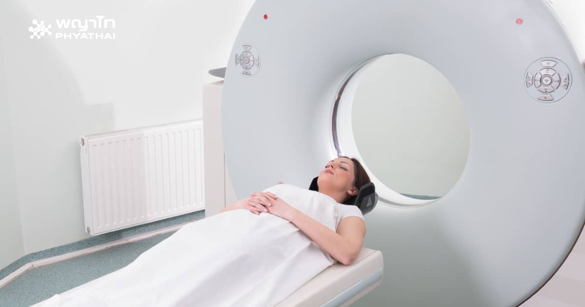
When is it necessary to have a brain scan?
If you have symptoms indicating a skull fracture, brain hemorrhage, loss of consciousness, a car accident, a fall from a height of more than 3 feet, or come to see a doctor with the following symptoms:
- Seizures
- Severe headache
- Frequent vomiting
- Decreased visual ability
- One pupil larger than the other
- Pain when pressing above the skull
- Problems with speech, hearing, and swallowing
- Fluid or blood coming from the ears and nose
- Weakness in the facial or one side of the body muscles
CT Scan is the best first choice that doctors will use to evaluate the cause of abnormal symptoms. However, if you have the above symptoms continuously for more than 48 hours after injury or if the symptoms worsen, MRI may be a better option.
How is MRI different from CT Scan?
Currently, the detection of abnormalities in various internal organs has advanced significantly. Both CT (computerized tomography) and MRI (magnetic resonance imaging) are widely used technologies. Although these two methods are very similar, there are several differences, which are:
- CT scan uses radiation, but MRI does not use radiation
CT scan uses x-ray beams passing through the patient’s body to create shadow images on a screen that receives the beam on the other side of the body. Meanwhile, MRI creates an electromagnetic field around the patient and detects changes in energy within that magnetic field without using x-ray beams. Exposure to such electromagnetic waves has not been found to affect health, so MRI is considered safer than CT scan.
- MRI is suitable for soft tissue examination, but CT is suitable for bone examination
MRI detects the movement of protons in water while in the magnetic field. Therefore, organs with high water content, such as soft tissues, muscles, blood vessels, brain tissue, etc., generate signals that can be detected well. Bones, which contain almost no water, cannot generate signals detectable by MRI. Therefore, for diagnosing bone-related diseases, CT scan should be chosen.
- CT takes less time than MRI
CT machines emit beams through the patient’s body while rotating around the patient. The rotation takes only 1-2 seconds to complete a full circle, producing a 360-degree image of the area. Thus, the total CT scan time usually does not exceed 10-15 minutes. MRI, on the other hand, takes much longer, sometimes up to 1 hour, which may be problematic for some patients who fear confined spaces (claustrophobia).
- The contrast media used are different
For CT scans, contrast agents containing iodine are injected to enhance image clarity. These iodine-based agents can potentially cause kidney toxicity, so special caution is needed when performing CT scans on patients with pre-existing kidney disease.
For MRI, the contrast agent used is Gadolinium, which does not contain iodine and therefore does not cause kidney toxicity. However, MRI contrast agents may cause a long-term skin disorder called Nephrogenic systemic fibrosis, especially in patients with kidney function measured by a glomerular filtration rate (GFR) less than 30 ml/min.
- Metal is prohibited for MRI
During MRI, the patient is placed inside a large magnetic field. If there is metal inside or outside the body within this magnetic field, it may move and cause harm. Therefore, most medical devices or metal implants in the body are contraindications for MRI. CT scans can be performed on patients with metal implants, but the images may be blurred because metal blocks x-ray beams, causing shadows near the metal.
However, nowadays, CT Scan and MRI are also used for screening to detect lesions early without waiting for abnormalities or adverse events to occur. This, of course, depends on the physician’s discretion as well.
