With the current social conditions, couples are marrying later, which also makes pregnancy more difficult and riskier regarding whether the fetus will be healthy and strong… How great would it be if we could check whether the baby to be born has any abnormalities? Because modern medical technology can diagnose these abnormalities while the fetus is still in the womb.
What causes fetal abnormalities?
Fetal incompleteness or abnormalities can occur due to
- Genetic risks, such as thalassemia, hemophilia, etc.
- Risks from advanced maternal age, such as Down syndrome, etc.
- Abnormalities that may occur spontaneously from abnormal fetal development during the embryonic stage, such as anencephaly, hydrocephalus, cleft lip and palate, etc.
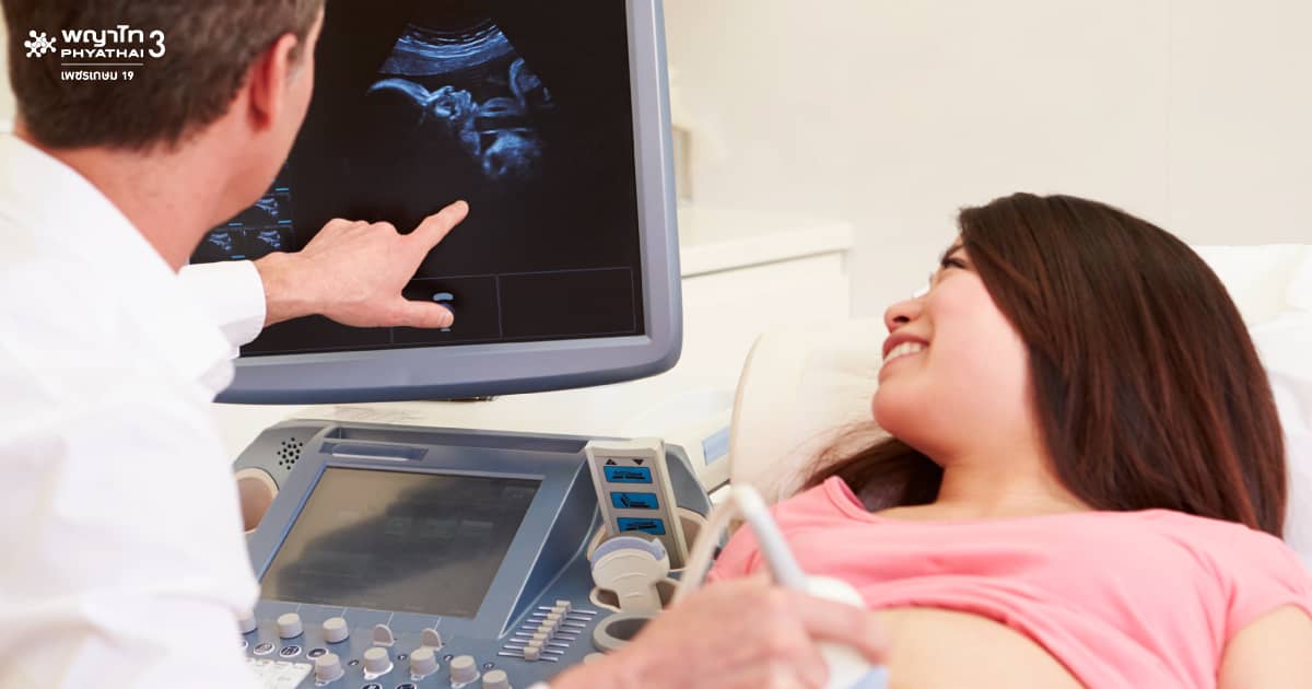
Check the body’s completeness with “Ultrasound”
Ultrasound examination helps diagnose the physical characteristics of the fetus well, such as the completeness of the head, arms, legs, and torso. This method of examination at different gestational ages provides different benefits as follows:
- First 3 months – Useful for calculating gestational age, determining intrauterine or ectopic pregnancy, whether the fetus is developing into an embryo, or detecting uterine fibroids and ovarian cysts during pregnancy more clearly than at later gestational ages.
- 4 to 6 months – The physical characteristics of the fetus’s major organs become clearer. If severe abnormalities are found, such as absence of the skull or an open abdomen, pregnancy termination may be considered immediately. If the fetus has a cleft lip or is significantly smaller than the standard, amniocentesis may be performed to check for chromosomal abnormalities to help decide whether to terminate the pregnancy.
- Last 3 months – Ultrasound is used to observe changes or growth of the fetus and whether the fetal organs are still intact. Some abnormalities may not be clear in the early stages, such as kidney and urinary tract abnormalities or hydrocephalus, to plan for diagnosis and treatment after birth.
There is no clear standard on how many ultrasounds should be done because it depends on the doctor’s assessment of each mother and the readiness of the healthcare facility.
Detect unseen abnormalities with chromosomal testing
The fetus may have no visible physical abnormalities. Chromosomal testing examines the genetic unit level, requiring fetal cell samples for analysis. Currently, fetal cells can be tested both before and after pregnancy.
- Pre-pregnancy stage – Used with in vitro fertilization, where embryonic cells before implantation into the mother’s uterus are tested for chromosomal abnormalities.
- Post-pregnancy stage – Used for mothers aged 35 and older, where amniocentesis is performed at 16-18 weeks gestation to collect fetal cells floating in the amniotic fluid for chromosomal abnormality testing. The most common abnormality found is Down syndrome, caused by an extra chromosome 21.
Gene abnormality testing from the embryonic stage
Gene abnormality testing examines the sequence of genetic units called “genes.” It can be done during in vitro fertilization or after pregnancy. Diseases that may be detected include genetic disorders such as thalassemia and hemophilia.
Diagnosis of fetal diseases is continuously developing and becoming more accurate, which will increase the possibility for couples who want fewer children but with strong and healthy offspring.
