Every expectant mother likely has the urge to sneak a peek at her baby in the womb at least once (or many times). Some may want to know if their baby is healthy and fully developed, while others are curious about what their baby is doing or simply want to see their little one’s face for reassurance. However, some mothers may still harbor concerns about whether frequent ultrasounds during pregnancy could harm their baby. They may wonder if such scans expose their child to harmful alpha or gamma radiation, or if there are any risks of brain defects or disabilities in the future.
Today, we aim to clarify any doubts and assure mothers that ultrasound examinations are beneficial for prenatal care and safe for both the mother and the baby.
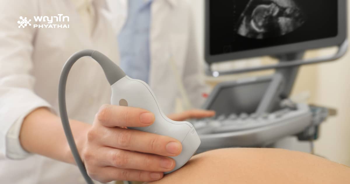
What is an Ultrasound?
Ultrasound (also known as a “high-frequency sound wave examination”) is a procedure that uses sound waves to create images of the baby in the mother’s womb. The equipment emits sound waves that travel through the mother’s body. These sound waves reflect off muscles, fat, abdominal organs, and the baby, placenta, and amniotic fluid within the uterus. The reflected waves return to the device, which converts them into images displayed on a screen.
You can think of it like the way dolphins or bats emit sound waves that bounce back to help them locate objects around them.
Why Do Mothers Need Ultrasounds?
The primary purpose of an ultrasound is to assess the health of the fetus. The benefits of ultrasound can be categorized based on the stage of pregnancy, as shown:
Summary of Ultrasound Examinations During Pregnancy
6-8 Weeks:
- Confirm pregnancy and number of fetuses; check for heartbeat.
- Assess risk for ectopic or molar pregnancy.
- Check for uterine fibroids or ovarian cysts that may affect pregnancy.
10-14 Weeks:
- Screen for severe congenital disabilities.
- Measure nuchal translucency to assess risk of Down syndrome.
18-22 Weeks:
- Perform detailed anomaly scan for fetal abnormalities.
- Assess fetal growth and weight.
- Examine placenta location, umbilical cord, and amniotic fluid volume.
28-36 Weeks:
- Reassess for fetal abnormalities before delivery.
- Monitor fetal growth and weight.
- Evaluate fetal health, including breathing and movements.
- Check placental health and amniotic fluid levels.
- Conduct Doppler studies for blood flow in umbilical cord and fetus.
- Confirm fetal position before delivery.
When Should Mothers Receive Ultrasound Examinations?
Mothers should have at least one ultrasound examination during the 18-22 week gestation period, performed by a maternal-fetal medicine (MFM) specialist, to check for fetal anomalies (anomaly scan) and to monitor fetal development. Current recommendations suggest that mothers should undergo at least one ultrasound in each trimester.
For mothers with complications such as low fetal weight, oligohydramnios, preeclampsia, diabetes, or multiple pregnancies, more frequent and detailed ultrasounds may be required. In some cases, ultrasounds may need to be conducted monthly, weekly, or even daily.
Is Frequent Ultrasound Dangerous?
Current data shows no evidence that ultrasound examinations pose any risks to mothers or fetuses regarding congenital disabilities, development, growth, cancer, psychosis, or other abnormalities after birth. Mothers can feel confident in undergoing ultrasound examinations.
Can Ultrasound Detect All Anomalies?
While ultrasound cannot detect every abnormality or disability, experienced specialists can typically identify about 80-90% of abnormalities. Limitations in detecting anomalies may include:
- Identifying specific organs affected (e.g., hands, feet, limbs, respiratory system) which can be difficult.
- Fetal positioning during the examination (e.g., lying face down, covering the face, crossing legs).
- Maternal body shape (obesity) or anomalies that develop later.
- Increased clarity of abnormalities that may be observed in later ultrasounds.
Therefore, mothers are advised to have at least one ultrasound examination per trimester (every three months).
Accuracy of Detecting Fetal Anomalies
The following shows approximate accuracy rates for detecting anomalies in various organs:
- Brain and Central Nervous System: 53-84% accuracy
- Heart / Severe Heart Defects: 9-13% / 50% accuracy
- Cleft Lip and Palate: 18-75% accuracy
- Abdominal Wall Defect: 80-98% accuracy
- Diaphragmatic Hernia: 50-58% accuracy
- Respiratory System: 20-50% accuracy
- Gastrointestinal System: 42-61% accuracy
- Urinary System: 57-70% accuracy
- Bones / Severe Bone Defects: 8-22% / 60% accuracy
- Arms, Legs, or Hands and Feet: 13-18% accuracy
