Cataract is a condition where the eye’s lens gradually becomes cloudy, which mostly occurs naturally. The risk increases after the age of 40. Today, we will introduce cataracts from various perspectives along with complete treatment through 5 different surgical methods.
What You Should Know About Cataracts
- There are more than one type of cataracts, but the most common type is cloudiness in the center of the lens. Less common types include cloudiness at the front, back, or edges of the lens.
- Children can also be born with cataracts, which may be caused by genetics, certain diseases, infections, or injuries before birth. Having cataracts from childhood significantly affects the development of the eyes and brain and may lead to vision loss.
- Diabetes is one of the factors that increase the risk of developing cataracts earlier and at a younger age. Diabetic patients have high blood sugar levels that cause the cells in the lens to become cloudy and reduce transparency, which can lead to cataracts causing blurred vision, faded images, or seeing images with a yellowish tint.
- After the cloudy lens is surgically removed, patients will not develop cataracts again because ophthalmologists insert a clear artificial lens in its place. Some patients may experience blurred vision after surgery due to the proliferation of lens epithelial cells remaining after cataract removal. In such cases, laser treatment can dissolve these cells, helping to restore clearer vision.
- People with severe nearsightedness have a high risk of retinal detachment after cataract surgery.
- Cataracts have affected the artwork of world-renowned artists such as Claude Monet, a French Impressionist painter famous for depicting changes in light, color, and shadow over time. Between 1912-1922, as Monet’s cataracts worsened, he saw changes in color intensity. Whites, greens, and blues he once saw clearly changed, resulting in larger brush strokes and more use of yellow, brown, and purple in his works.
- Iris color affects the risk of developing cataracts. Studies have found that people with dark brown irises have a higher chance of cataracts than those with lighter-colored irises. UV light is a factor that can cause cataracts regardless of iris color. Therefore, wearing sunglasses to protect against UV rays and wide-brimmed hats when outdoors can help reduce the risk of cataracts.
- In some cases, ophthalmologists may recommend cataract surgery on both eyes, even if one eye still sees well. However, most patients do not have surgery on the second eye until the cataract affects daily life. Surgery with artificial lenses in both eyes helps balance vision and reduces the risk of falls that can cause bone fractures. Additionally, implanting an artificial lens in the second eye can reduce the risk of angle-closure glaucoma in some patients later on.
Cataract Treatment by Surgery
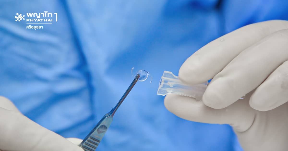
Cataracts can only be completely cured by surgery. Before surgery, anesthetic eye drops or injections are given to ensure the patient does not feel pain during and after the procedure. General anesthesia is used only in certain cases. The surgery consists of two main steps:
- Removing the cloudy natural lens, and
- Inserting a clear artificial lens in the same position to restore normal vision.
5 Surgical Methods for Cataract Treatment
1. Cataract Surgery by Phacoemulsification
Phacoemulsification surgery involves a small incision of about 2.2-3.0 millimeters. The ophthalmologist inserts instruments through the incision and uses high-frequency ultrasound waves to break up the cataract and suction it out, leaving the lens capsule intact to place the artificial lens. This method usually requires no stitches or only minimal stitching because the small incision is strong, self-sealing, and heals quickly. It causes less irritation, less astigmatism from surgery, and allows faster visual recovery.
2. Laser-Assisted Cataract Surgery (Femtosecond Laser-Assisted Cataract Surgery)
This technology makes cataract surgery easier. The femtosecond laser system for cataract surgery was first used in 2011 and assists in several surgical steps, including:
- Creating the corneal incision as an entry point for surgical instruments
- Opening the front lens capsule
- Fragmenting the cataract into small pieces for easier emulsification and removal
- Making corneal relaxing incisions to correct refractive errors in some cases
Although femtosecond laser surgery is precise and safe, it is more expensive due to advanced technology. Moreover, the postoperative results are similar to phacoemulsification, which also uses a small incision.
3. Extracapsular Cataract Extraction (ECCE)
This method is used when the cataract is very dense and hard, making removal through a small incision impossible. The incision is about 8-10 millimeters wide to remove the cataract in one piece, leaving the lens capsule intact to insert the artificial lens, followed by suturing the wound closed.
4. Manual Small Incision Cataract Surgery (MSICS)
This technique removes the cataract in one piece through a small self-sealing incision in the sclera without sutures, leaving the lens capsule in the eye. The incision is smaller than ECCE but larger than phacoemulsification. The surgical outcomes are comparable to phacoemulsification. It is a good alternative for developing countries because it requires less surgical time and is less expensive. Additionally, no sutures reduce the risk of postoperative astigmatism, speed up recovery, and reduce the number of follow-up visits.
5. Intracapsular Cataract Extraction (ICCE)
This method is used only in very specific cases.
Post-Cataract Surgery: Monitor for Abnormalities
If the patient has no other eye diseases, the optic nerve and brain controlling vision are normal, and there are no surgical complications, vision will improve after surgery. Therefore, if vision does not improve or becomes blurry after surgery, consult an ophthalmologist to identify the cause and receive further treatment.
