At present, the incidence of cancer in Thailand has been increasing, which is also found that cancer is a number one non-communicable diseases causing death to people
A number of patient with cancer is rising higher, that is caused by many factors. Such as environment, habits, and consumption. However, the reason that cancer incidence is found due to doctors are able to diagnose and detect more accurate and faster. A vital support comes from an advanced radiology technology. It supports a diagnosis process, including helping physicians detect the distribution of the disease more accuracy and clear. For that reason physicians can accurately identify the stage of cancer and provide a good solution for a team of cancer specialists (Multidisciplinary Team: MDT) However recently, the radiologic technology that help diagnosis the cancer are as following:
1. Digital Mammogram Machine
It is a special x-ray machine for breast examination, which is more effective than a mammogram film. Patients will receive low radiation with an accurate 90% result. And it will distinguish the difference of fat and the breast tissues clearly. Including, abnormalities, position of the tumor and pathogen, which can be seen in 1 cm. accordingly, cancerous tissue in the breast duct is only be seen when it is at the beginning of the transitionm also known as DCIS (ductal carcinoma in situ) where the cancer cannot be found. Most of cancer demonstrates in the form of clustered of microcalcifications. Moreover, breast cancer has become common diseases for females in Thailand, which found increasing more numbers of patient in every year. There are about 30% of people who are diagnosed with breast cancer at an early stage. If receive a surgery and treatment follow a physician direction that will help providing better results. And also improve a life quality of breast cancer patient.

Figure 1 Breast cancer in the early stages may be characterized by microcalcification, ill-defined mass spiculated margin, distortion, knobby, lobulated or smooth contour mass in 90% breast sensitivity and specificity of 95%
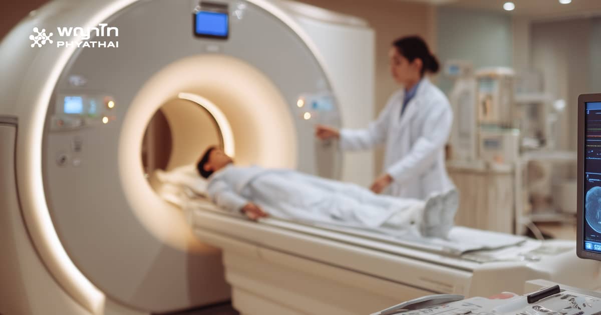
2. A computerized tomography (CT Scan) or MDCT Scan a machine which is developed with modern technology including high speed and resolution. Creating 64 images at one time rotation or scanning 360 degrees in 0.4 seconds, resulting in 2,000 images. Accordingly, the results have a high accuracy such as some particular organs like the heart. Which in general a computer x-ray machine usually results in a low image resolution. But, the CT scan can produce clear images for both 2D and 3D, as well as all parts of the body such as veins, tissues, bones, heart or including trachea in the lung area.
These are reasons why the computerized tomography is beneficial for diagnosis and treatment plan. Including therapeutic planning which precisely predicts a severity of cancer. And also detect abnormalities of the large intestine with a new technique called Virtual Colonoscopy that can detect small lesions which may become a cause of colon cancer.
At present colon and anal cancer are found to be more increased due to lifestyle change. From the latest cancer statistics in Thailand, was found that if combine a number of people from both diseases in males and females, that can be more than a number of those who have cervical cancer. However, CT scan also helps detecting lung cancer in the first stage. Due for lung x-ray the stage may not be seen, this makes a treatment even more effective and has more opportunities for a cure as well.
3. MRI (Magnetic Resonance Imaging) a visualized machine used for disease diagnosis. Which requires a magnetic property of hydrogen atom inside the body under the Electromagnetic fields. Accordingly, that can detect abnormalities in different organs within the body from the initial stages ofdisease, for example the stage that occurs in the brain, spine, abdominal cavity organs, pelvic and breast called (MR mammography) Including bile duct, gallbladder (MRCP) and musculoskeletal system. That also include veins and arteries (MRA, MRV), special examinations as MR Spectroscopy. The examination used to detect biochemical aspect in tissues in order to remove tumors from normal tissue. Function MRI , this is an inspection for the organs functioning.
Breast examination with MR mammography currently, the MRI is recommended to do with a mammogram. Especially for those who have been diagnosed with BRCA, gene mutation by the age of 30 or more. Including, who have had a high dose radiation around the chest area, since a young age and an inspection of lesion that may be found more than one location. Also, to follow up symptoms after oncoplastic breast surgery in order to classify between cancer and fascia.
4. SPECT or single photon emission computed tomography is a nuclear medicine used for disease diagnosis and it can be inserted in the body for organ inspection. However, a principle of a process is to inject substances called Radiopharmaceutics. These substances can get into the body’s systems, such as bones that have Inflammation or degeneration. Meaning some of Radiopharmaceutics inspect the kidney and liver function. Some get into to the digestive tract where there is bleeding. And some substances monitor the thyroid gland. Later, a doctor will apply Gamma Scanner or gamma camera for radiation scanning. And displays the organs images afterward, also demonstrates about their works and functions. In addition, to provide the Radiopharmaceutics , a nurse will inject a small amount of them through the arm vein. However, they are safe for the body. After done with injection, you may wait for a while. Then go to bed where there is a gamma camera scanning and lay down, a duration takes about 20 – 40 minutes. Which a doctor diagnose symptoms from images.
5. PET Scan: Positron Emission Tomography Scanner it is a diagnostic tool of nuclear medicine for cell’s metabolism inspection. Which detects a degeneration and death of cells, including rapid cells division, by using Radiopharmaceutics obtained from cyclone, inject to patients and measure a radiation intensity from lesion according to a level of cells function. For example, quick division where cancer cells absorb more Radiopharmaceutics than normal cells. The scanner will detect radiation and shot pictures. That is beneficial to diagnose whether the image of lumps seen x-ray is cancerous or not. Including, assessing spread of cancer, a response to cancer treatment, and authentic prognosis.
In addition, PET scan can also detect abnormality of nerve system as well as cardiac function. For Positron emission tomography or Computed tomography, it is an innovation of nuclear medicine and computertomography combined together. PET will provide a visual representation of biological and metabolic processes. While CT will show anatomical information as what size, shape, and position of pathology are in the body. However, integration of anatomical and metabolic pictures (co-registration) helpsproviding a location of pathology accurately. Including, telling a location where the radiopharmaceutical is, known as physiologic uptake, also pathological location or pathological uptake. Accordingly these two ways assist a doctor in diagnosis stage of cancer, both for staging and restaging. That also include prognosis, treatment evaluation, recurrence of cancer analyzing, and the treatment reaction.
Beside, radiation therapists will use PET / CT images for radiation therapy treatment planning. Which can accurately determine the position of radiation or contour maps and it can cover the lesions as well. However, receiving PET can be expensive, fast and less than 100 percent of accuracy. Thus, PET examination cannot be used to cover all of examinations. But only be one ofadditional inspections for cancer which increases more authentic results after receiving standard examinations, such as mammogram, CT or MRI. Also helpcreating more accuracy for cancer stage classification, which support a next treatment provided by doctor.
6. Ultrasound, it is a diagnostic tool that people commonly know and receive to examine the internal organs of the body with high frequency sound waves that cannot be heard, therefore, there is no harm. As that reason, this type of sound waves is used to detect almost in every organ and tissue inside the body. Except some organs contain gas or solid such as bones, lung, bowel, digestive tract. Due to high-frequency reflection is unable to produce an image with a good resolution. Moreover, technically the advantages of ultrasound are safety, fast, convenient, pain-free and having multiplanar for each examination. This is used to observe the cancer distribution to the liver, including the size and position of the lumps. For that reason, it is also used to support mammogram in order to see details of cancerous tumors and find new blood vessels (neovascularization) that support the lumps also assess an elasticity of the tumor. However, connective tissues or elastogram is also included.
Cancer caused by a lifestyle changing that has a high incidence of cancer being diagnosis with radiation for cancer is an important step for identifying the disease and detecting a severity so that physicians can provide an appropriate treatment to patients. An early detection is more useful due to cancer still can be cured in the early stage.

Phyathai1 hospital
